Cerebrovascular Surgery
Giving you the
very best
of care.
Cerebrovascular disorders involve treating the blood vessels of the brain and spinal cord and require a highly specialized expertise.
As an experienced and highly skilled neurosurgeon, Dr. Ehtesham is well-versed in treating numerous cerebrovascular disorders. Even though there is a lot of fear surrounding words like stroke, aneurysm, and arteriovenous malformations, Dr. Ehtesham will be your trusted guide through the process. He will not only provide you with expert care along your journey but will help ease your fears through information, education, and communication.


Brain
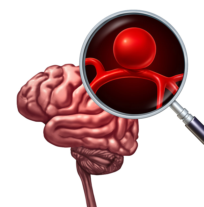
Aneurysms
A cerebral or intracranial aneurysm is a bulge or ballooning of an arterial blood vessel in the brain. If an intracranial aneurysm ruptures, it can result in stroke, brain damage, or death. That’s why it’s so important to seek immediate treatment if you experience symptoms such as:
- An unusually severe headache
- Nausea
- Stiff neck
- Sensitivity to light
Most intracranial aneurysms occur between the underside of the brain and the base of the skull. A ruptured aneurysm quickly becomes life-threatening and requires prompt medical treatment. However, most brain aneurysms that don’t rupture still create health problems or cause symptoms. Such aneurysms are often detected during tests for other conditions.
An unruptured aneurysm usually causes no symptoms. A key symptom of a ruptured aneurysm is a sudden, severe headache.
Treatment
Dr. Ehtesham is one of very surgeons trained in how to treat brain aneurysms with both open surgery and minimally invasive endovascular techniques. He primarily uses one of three different techniques to treat brain aneurysms, depending on whether the aneurysm is ruptured or unruptured, and where it is located.
Endovascular embolization, or coiling, halts the flow of blood into the aneurysm by sealing it off. A relatively new and minimally invasive procedure, it has become the treatment of choice. A catheter is inserted into an artery in the patient’s leg and guided to the aneurysm. Tiny platinum coils are threaded through the catheter and released to fill the entire aneurysm so blood flow into it is stopped.
Flow diversion is a technique Dr. Ehtesham uses to divert the flow of blood away from the aneurysm by placing a stent (a soft, flexible mesh tube) inside the blood vessel where the aneurysm is located. In time, new cells grow on the stent, sealing the aneurysm and healing the blood vessel.
Microsurgical Clipping is a common surgical treatment. A craniotomy is performed to open a section of the skull so that Dr. Ehtesham can access the aneurysm beneath. A tiny metal clip is attached to the base of the aneurysm so blood flows past it. This helps prevent possible rupture or re-rupture.
Strokes
A stroke occurs when the blood supply to part of your brain is interrupted or reduced, preventing brain tissue from getting oxygen and nutrients. Brain cells begin to die in minutes.
Most strokes are caused by an abrupt blockage of arteries leading to the brain (called schematic stroke). Other strokes are caused by bleeding into brain tissue when a blood vessel bursts (called hemorrhagic stroke). Because strokes appear rapidly and require immediate treatment, a stroke is also called a brain attack. When the symptoms of a stroke last only a short time (less than an hour), this is called a transient ischemic attack (TIA) or mini stroke.
A stroke is a medical emergency, and prompt treatment is crucial. Early action can reduce brain damage and other complications.
Symptoms of a stroke can include:
- Trouble speaking or slurring your words
- Understanding what others are saying and general confusion
- Paralysis or numbness of the face, arm, or leg
- Vision problems
- Sudden, severe headache
- Vomiting, dizziness, or altered consciousness
- Trouble walking or loss of balance
Treatment
Early treatment with medications like tPA (clot buster) can minimize brain damage. Other treatments focus on limiting complications and preventing additional strokes.
Some stroke victims may benefit from surgery that is designed to remove or dissolve blood clots and stop bleeding in the brain. Dr. Ehtesham performs several different procedures to help, depending on what type of stroke the person has suffered.
If the stroke has been caused by a blockage in the carotid artery in the neck – which supplies blood to the brain – Dr. Ehtesham may recommend a minimally procedure known as a carotid endarterectomy to remove the buildup of plaque in the artery. In this procedure, he makes a small incision in the neck, opens the artery, removes the plaque, then closes. The procedure is typically performed using a local anesthesia, with an overnight hospital stay for observation.
If an aneurysm caused the stroke, Dr. Ehtesham typically recommends one of two procedures:
Endovascular embolization, or coiling, halts the flow of blood into the aneurysm by sealing it off. A relatively new and minimally invasive procedure, it has become the treatment of choice. A catheter is inserted into an artery in the patient’s leg and guided to the aneurysm. Tiny platinum coils are threaded through the catheter and released to fill the entire aneurysm so blood flow into it is stopped.
Microsurgical Clipping is a common surgical treatment. A craniotomy is performed to open a section of the skull so that Dr. Ehtesham can access the aneurysm beneath. A tiny metal clip is attached to the base of the aneurysm so blood flows past it. This helps prevent possible rupture or re-rupture.


Arteriovenous
Malformations
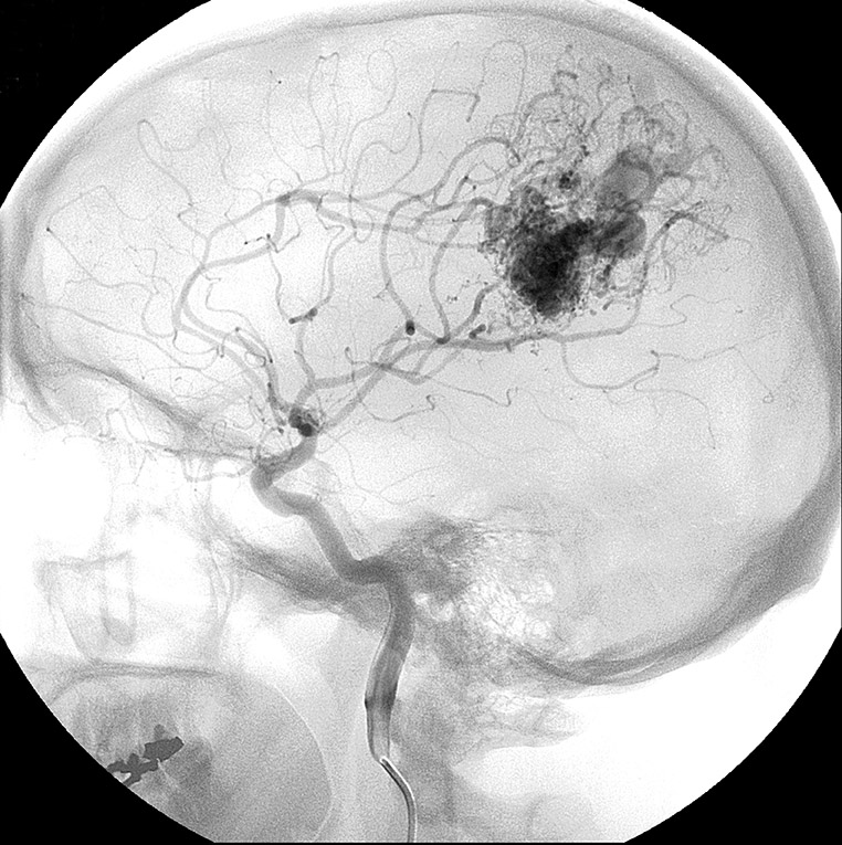
An arteriovenous malformation (AVM) is a rare defect of the circulatory system that develops prior to birth or soon after. Blood flows from an artery directly into a vein through a passage known as a fistula, which deprives the surrounding tissue of oxygen.
Although an arteriovenous malformation can occur anywhere in the body, it can have serious consequences when it occurs in the brain or spinal cord where besides reducing the amount of oxygen delivered to the brain, it may press on nerves or cause hemorrhaging.
The most common symptom of an AVM is headaches. Other symptoms include hemorrhaging (bleeding), seizures and other neurological problems such as paralysis, muscle weakness, and loss of speech, vision, coordination, or memory. Only about 12 percent of people with AVMs have symptoms. Symptoms are most often noticed in a person’s twenties, thirties, or forties.
Treatment
The main goal of AVM treatment is to prevent hemorrhage, but a strong secondary goal is to control any seizures or other neurological problems that may have manifested.
Not all arteriovenous malformations need surgery but when surgery is necessary, Dr. Ehtesham may use conventional surgery to remove the fistula. Where possible, he will then use a less invasive approach to plug the fistula. One of the less-invasive procedures Dr. Ehtesham uses is called an endovascular embolization. Considered an alternative to open surgery, endovascular embolization (EE) blocks blood vessels in order to cut off blood flow to the affected area. After making a tiny incision in the groin, Dr. Ehtsham inserts a long, thin tube, called a catheter, through the femoral artery. The catheter is guided through the circulatory system using X-ray imaging until it reaches the location of the abnormal blood vessel being treated. He then injects an embolizing agent, such as tiny plastic particles, metal coils, or glue-like agents, to block the artery and reduce or stop the blood flow.

